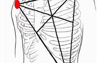
图10 应用Lewis导联后可记录到清晰的P波
如此看来,无意为之的导联位置错误可能干扰诊断,而有意为之的导联位置改变却可能有事半功倍之效。
心电图的乐趣和难点亦在于此!
参考文献:
[1] Francis J. ECG monitoring leads and special leads. Indian Pacing Electrophysiol J. 2016; 16(3): 92-95.
[2] Rodrigues de Holanda-Miranda W, Furtado FM, Luciano PM, Pazin-Filho A. Lewis lead enhances atrial activity detection in wide QRS tachycardia. J Emerg Med. 2012; 43(2): e97-9.
[3] Abobaker A, Rana RM. V1 and V2 pericordial leads misplacement and its negative impact on ECG interpretation and clinical care. Ann Noninvasive Electrocardiol. 2021 Apr 4: e12844.
[4] 郭继鸿. Brugada综合征的诊断与治疗. 临床心电学杂志, 2005; 14(3): 215-223.
[5] 孙海燕, 于小林, 孙迎春. 图解肢体导联电极错接的心电图表现. 江苏实用心电学杂志. 2013; 22(5): 827-829.